Tahıllarda Septoria hastalığı (Septoria tritici)

Tahıllarda Septoria hastalığı
Perfect dönemi: Mycosphaerella graminicola
Bitkinin gelişme evresine ve çevre şartlarına bağlı olan, bitkinin toprak üstü aksamını etkileyen ve değişik tipte leke oluşturan primer patojenlerdir (hastalık etmenidir).
Septoria hastalıklarını hububattaki diğer hastalıklardan şu özellikleri bakımından ayırabiliriz:
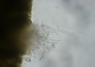
- Az veya çok küresel pycnidium denilen çoğalma organlarının içerisinde picniosporlar oluşur.
- Pycnidiumlar bitki dokularında gelişir. Bunlar olgunlaştıkça epidermisi patlatır. Ostiol denilen açıklıklardan veya deliklerden pycniosporlar toplu olarak dışarıya salınırlar. Bu spor kümeleri genellikle hafif pembe sarı renktedir.
- Pycniosporlar ipliksi ve silindirik şekilli, türe bağlı olarak boyutları değişebilir. Uç kısımları hafif kıvrık ve küttür. Renksiz, şeffaf ve 2-4 bölmeye sahiptirler.
Konukçuları
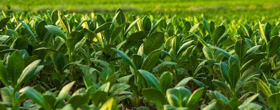
Esas olarak buğdayda parazittir; fakat uygun şartlarda triticale ve çavdarda, nadiren de arpa ve bazı yulaf türlerinde görülür.
Yayılma Alanları
Hastalık buğday yetiştirilen tüm ülkelerde görülür.
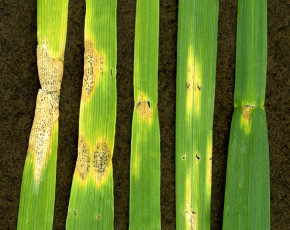
Belirtileri nelerdir ?
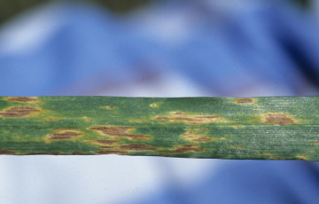
- Lekeler başlangıçta yaprak damarlarınca sınırlandırılan küçük, düzensiz, kırmızımsı, kahverengi şekilli olurlar.
- Lekeler damarlar boyunca uzunlamasına gelişmesine devam ederler.
- Hastalık ilerledikçe lekelerin rengi kül rengini alır, lekeler birleşerek tüm yüzeyi kaplar.
- Sonuçta tüm yaprakta nekroz meydana gelir.
- Lekeler büyürken etraflarındaki siyah renk halkası kaybolur ve parlak grimsi bir renk alırlar.
- Daha sonra bu lekelerin üzerinde küçük siyah noktacıklar görülür.
- Bu noktacıklar fungusun pycnidiumlarıdır.
- Hastalık genellikle bitkinin en alt yapraklarında görülür.
- Üst yapraklara yayılma oranı çevre şartlarına ve çeşidin çevre şartlarına karşı duyarlılık reaksiyonuna bağlıdır.
- Bitkiler olgunlaşmaya başladığında etmenin agresifliği azalmaya başlar.
- Hastalık ilerledikçe yaprak dokularında gömülü vaziyette çoğalma organları oluşmaktadır.
- Daha ileri devrelerde pycnidiumlar epidermisi patlatır, kahverengi-sarı renkli (jel gibi) spor kümecikleri dışarıya salınırlar.
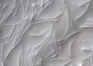
Spor Yapısı nasıldır ?
Pycnidiosporlar iplik benzeri (filiform) renksiz, çubuk şekilli, genellikle kıvrık, uçları yuvarlakça, çok belirgin olmayan 3-8 bölmeye sahiptir.
Kışlaması nasıl olmaktadır ?
Etmen kışı hastalıklı bitki artıklarında miselyum şeklinde geçirir.
Hastalığı ilkbaharda yaprak üzerinde oluşan pycnidiumlardan çıkan pycniosporlar başlatır.
Hastalık hakkında tanımlayıcı fotoğraflar aşağıda verilmiştir. Fotoğraflar Valent USA corporation, Purdue University, Montana State University tarafından telif haklarına sahiptir. İzinsiz çoğaltılamaz,yayınlanamaz, herhangi birde kullanılamaz.


English version
The initial symptoms of STB are small chlorotic spots on the leaves that appear soon after seedlings emerge in the fall or spring. As they enlarge, the lesions (Figure 2) become light tan and develop darker colored fruiting bodies (Figure 3). Lesions on mature leaves most often are long, narrow and delimited by leaf veins but also can be shaped irregularly or can be elliptical, particularly on seedlings or leaves that were young when infected.
Mature lesions contain black or brown fruiting structures, the asexual pycnidia or sexual pseudothecia. The pycnidia or pseudothecia develop in the substomatal cavities of the host so are spaced regularly within the lesions STB is found commonly in the same fields and on the same plants as Phaeosphaeria nodorum(asexual stage: Stagonospora nodorum), the causal agent of Stagonospora nodorum blotch of wheat. When both pathogens occur together, they are referred to collectively as the Septoria blotch complex or Septoria complex.
Disease cycle
Infection by M. graminicola is initiated by air-borne ascospores and splash-dispersed conidia produced on residues of the previous season’s crop. Primary infection occurs soon after seedlings emerge in fall (for winter wheat) or spring. Ascospore germ tubes are attracted to the stomata, through which they gain entry into the sub-stomatal cavity either directly or after production of an appressorium-like structure (infection cushion). For several days the hyphae grow intercellularly with little increase in biomass.
After the switch from biotrophic to necrotrophic growth, cells collapse, lesions form and are identified initially by small, yellow flecks or blotches. The lesions expand, primarily in the direction of the leaf veins to form long, narrow, necrotic blotches. Pycnidia develop around stomata within the necrotic areas of the lesions and exude conidia in gelatinous, hygroscopic cirrhi. These spores are disseminated by rain splash to leaves of the same or nearby plants. The pathogen survives crop-free periods primarily as pseudothecia but also in pycnidia on crop debris. Autumn-sown crops and volunteer plants can aid survival over winter.

Septoria tritici – life cycle
Epidemiology
Initial inoculum usually consists of airborne ascospores, which cause the primary infections on seedling leaves, but also can be from conidia. Primary infections from an ascospore shower will occur evenly over a crop and give rise to lesions that bear pycnidia, the asexual structures that allow for rapid dispersal of the secondary inoculum, conidia.
Secondary spread of STB is by conidia, which form readily in high humidity, particularly if there is free water present on the leaves, but also can be by ascospores. Pycnidia with conidia are produced roughly 14 to 40 days after infection, depending on the host and seasonal conditions. These spores disperse through rain wash and splashing, causing local spread of the disease to uninfected leaves of the same and nearby plants. Production and dispersal of conidia occurs quite rapidly compared to pseudothecia with ascospores, which take several weeks until ripening. Thus both conidia and ascospores contribute to the epidemic but the asexual cycle seems to dominate during the growing season.
Ascospores can be airborne over large distances, while conidia are unlikely to travel far from their site of origin by rain-splash dispersal. Conidia help to spread the disease upwards through the canopy. Infection of flag leaves (last leaf to emerge on a wheat stalk) is common and leads to greatly reduced yields and poor quality of harvested grain. Rain splash of conidia can lead to disease foci, which can give a patchy appearance to the overall disease distribution in a field. A more uniform appearance of the disease is typical when the airborne ascospores are plentiful during the initial infection.
Many cycles of sexual and asexual reproduction during the growing season allow epidemics to develop rapidly. Debris from heavily infected leaves and stems remains in fields after harvest to produce inoculum for the next growing season.
Selected References
Alisa Ponomarenko1, Stephen B. Goodwin2, and Gert H. J. Kema3
1Department of Botany and Plant Pathology, Purdue University, West Lafayette, IN
2USDA-ARS, Crop Production and Pest Control Research Unit, Purdue University, West Lafayette, IN
3Plant Research International, Wageningen,? The Netherlands
Agrios, G.N. 2005. Plant Pathology, 5th edition. Academic Press, Inc, San Diego. 922 pp.
Bockus, W.W., R.L. Bowden, R.M. Hunger, W.L. Morrill, T.D. Murray, and R.W. Smiley. 2010. Compendium of Wheat Diseases and Pests: Third Edition. American Phytopathological Society, St. Paul, MN
Goodwin, S.B. 2007. Back to basics and beyond: increasing the level of resistance to Septoria tritici blotch in wheat. Australasian Plant Pathology 36: 532?538.
Goodwin, S.B., C. Waalwijk, and G.H.J. Kema. 2004. Genetics and Genomics of Mycosphaerellagraminicola: a model for the Dothideales. Pages 315-330 in: Applied Mycology and Biotechnology. Volume 4, Fungal Genomics. Elsevier Press, San Diego.
Eyal, Z., A.L. Scharen, J.M. Prescott and M. van Ginkel. 1987. The Septoria diseases of wheat: Concepts and methods of disease management. CIMMYT. Mexico, D.F.
Kema, G.H.J., D.Z. Yu, H.J. Rijkenberg, M.W. Shaw and R.P. Baayen. 1996. Histology of the pathogenesis of Mycosphaerella graminicola in wheat. Phytopathology 86: 777-786.
Kema, G.H.J., J.G. Annone, R. Sayoud, C.H. Silfhout, M. van Ginkel, and J. de Bree. 1996. Genetic variation for virulence and resistance in the wheat-Mycosphaerella graminicola pathosystem I. Interactions between pathogen isolates and host cultivars. Phytopathology 86: 200-212.
Kema, G.H.J., R. Sayoud, J.G. Annone, C.H. Silfhout. 1996. Genetic variation for virulence and resistance in the wheat-Mycospharella graminicola pathosystem II. Analysis of interactions between pathogen and host cultivars. Phytopathology 86: 213-224.
Joint Genome Institute, United States Department of Energy and University of California.Mycosphaerella graminicolav2.0 ? Home http://genome.jgi-psf.org/Mycgr3/Mycgr3.home.html
Yazılarımızdan haberdar olmak için
Abone olmak istermisin ?
Kaliteli yazılarımız ve fotoğraflarımızdan ilk sizin haberiniz olsun !
Bitki koruma ailesine katıldığınız için teşekkür ederiz.
Bir şeyler yanlış gitti sanırım 🙁